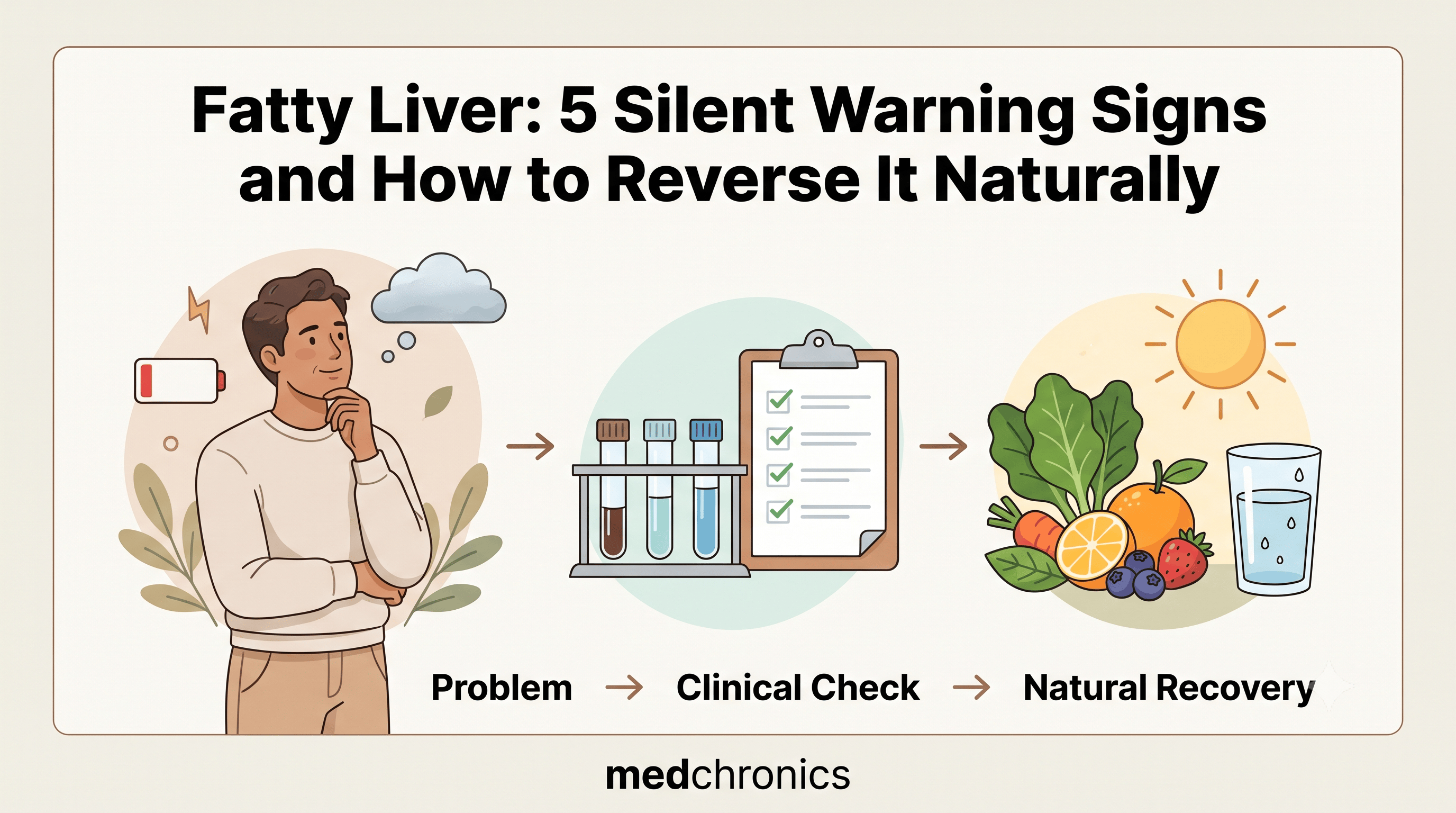
Fatty liver develops when excess triglycerides accumulate inside hepatocytes, silently compromising hepatic architecture and metabolic function long before clinical symptoms emerge. Known clinically as metabolic dysfunction-associated steatotic liver disease (MASLD, formerly NAFLD), this condition now impacts more than 30% of the global adult population. Because hepatic parenchyma lacks sensory pain receptors, significant intrahepatic lipid pooling often progresses undetected until advanced stages.
Table of Contents
Understanding Fatty Liver: Root Causes and Pathophysiology
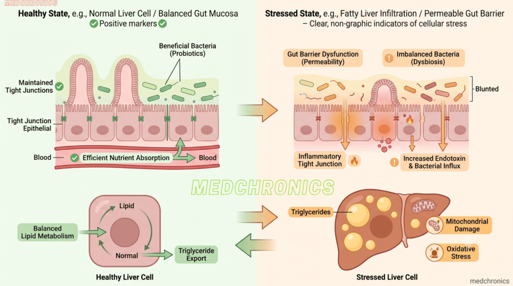
Hepatic steatosis (fatty liver) is primarily an endocrine and metabolic failure rather than an isolated hepatic disorder. When metabolic pathways become overwhelmed, the liver shifts from an efficient metabolic processing hub into an ectopic storage organ for excess lipid substrates.
- Insulin Resistance and Hyperinsulinemia: Chronic elevated circulating insulin prevents adipose tissue lipolysis from shutting down properly. This flood of non-esterified free fatty acids circulates directly into the portal vein and infiltrates liver tissue.
- De Novo Lipogenesis (DNL): High dietary intakes of refined carbohydrates and high-fructose corn syrup bypass normal hepatic energy checkpoints. Fructose is metabolized exclusively in the liver, converting surplus caloric energy directly into new intrahepatic triglycerides.
- Mitochondrial Overload and Oxidative Stress: As hepatic cells become saturated with lipids, mitochondrial beta-oxidation falters. Incomplete fatty acid oxidation generates reactive oxygen species (ROS), triggering hepatocyte ballooning, inflammatory cytokine release, and progressive fibrosis.
5 Silent Warning Signs of Fatty Liver Disease
Because early hepatic steatosis (fatty liver) rarely presents with acute pain, identifying subtle metabolic and systemic cues is critical for timely intervention.
1. Persistent Postprandial Fatigue and Brain Fog
Mitochondrial energy production declines when hepatocytes are congested with lipid droplets. Patients often report profound, unrefreshing sluggishness and cognitive haze, particularly 60 to 90 minutes after carbohydrate-dense meals. This systemic exhaustion reflects impaired hepatic gluconeogenesis and circulating inflammatory cytokines like TNF-alpha.
2. Right Upper Quadrant Fullness or Dull Ache
The liver parenchyma itself has no nociceptive nerve fibers, meaning fat buildup itself does not hurt. However, as progressive hepatic enlargement (hepatomegaly) occurs, the organ stretches its fibroelastic covering, known as Glisson’s capsule. This anatomical stretching triggers a chronic, vague fullness or dull heaviness beneath the right lower ribcage.
3. Cutaneous Markers: Acanthosis Nigricans and Skin Tags
Cutaneous manifestations often mirror internal hepatic insulin resistance.
- Acanthosis Nigricans: Dark, velvety hyperpigmentation localized to intertriginous zones such as the posterior neck, axillae, and groin folds.
- Acrochordons (Skin Tags): Multiple small, benign fibroepithelial polyps erupting around the neck and torso, driven by hyperinsulinemia-induced epidermal proliferation.
4. Visceral Adiposity Combined with Dyslipidemia
A expanding waist circumference (greater than 40 inches in men, or 35 inches in women) strongly correlates with visceral fat enveloping internal organs. When accompanied by an elevated triglyceride-to-HDL ratio (greater than 3:1), this clinical picture reliably indicates saturated adipose storage capacity and active fat spillover into the hepatic parenchyma.
5. Fluctuating or Borderline Liver Enzymes (ALT and AST)
Mildly elevated Alanine Aminotransferase (ALT) and Aspartate Aminotransferase (AST) levels frequently signal active hepatocyte stress or micro-necrosis. Importantly, advanced steatosis can exist even with normal transaminase values; an elevated serum ferritin level or an ALT/AST ratio greater than 1 often warrants deeper elastography or ultrasound investigation.
How to Reverse Fatty Liver Naturally: Evidence-Based Strategies
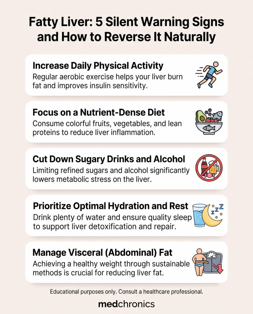
The liver possesses remarkable regenerative capacity. When lipotoxic stress and hyperinsulinemia are systematically removed, hepatocytes can eliminate accumulated fat droplets and resolve early-stage fibrosis.
Eliminate Liquid Fructose and Industrial Refined Sugars
Liquid sugars are the fastest dietary route to hepatic steatogenesis. Fructose circumvents phosphofructokinase, flooding hepatic glycolysis and initiating unchecked lipogenesis.
- Cut liquid sweeteners: Completely eliminate sodas, sweetened iced teas, commercially bottled fruit juices, and energy drinks.
- Read packaged labels: Minimize products listing high-fructose corn syrup, agave nectar, maltodextrin, and invert sugar among the first five ingredients.
- Prioritize whole fruit: Consume whole, fiber-rich berries, apples, and citrus rather than blended smoothies or juices to blunt glucose absorption.
Adopt an Anti-Inflammatory Mediterranean Pattern
The Mediterranean dietary template consistently outperforms low-fat protocols for reducing intrahepatic lipid pools and improving systemic insulin sensitivity.
- Emphasize monounsaturated fats (MUFAs): Use extra virgin olive oil as your primary fat source to preserve cell membrane fluidity.
- Increase omega-3 polyunsaturated fats: Consume wild cold-water fish (mackerel, sardines, wild salmon) twice weekly to attenuate hepatic inflammation.
- Pack in cruciferous vegetables: Incorporate broccoli, Brussels sprouts, and arugula, which provide sulforaphane to stimulate phase II liver detoxification pathways.
Deploy Time-Restricted Eating (TRE)
Limiting caloric consumption to a daily 8 to 10 hour window forces your metabolism to transition from glucose burning to fat oxidation. Extended fasting intervals deplete hepatic glycogen stores, stimulating macroautophagy—the cellular clean-up mechanism through which hepatocytes degrade internal lipid droplets. Start with a structured 14:10 fasting protocol before progressing to a 16:8 regimen.
Integrate Concurrent Aerobic and Resistance Training
Exercise rapidly decreases liver fat even in the absence of total body weight loss. Muscle contractions act as an endocrine sink, clearing glucose and circulating free fatty acids before they can enter the portal circulation.
- Zone 2 Cardio: Perform 150 minutes weekly of low-intensity aerobic work (conversational pace) to maximize mitochondrial fat oxidation.
- Progressive Resistance Training: Perform full-body compound lifts or bodyweight exercises 3 days weekly to upregulate peripheral GLUT4 transporters.
Utilize Targeted Hepatoprotective Nutrients
Certain clinical micronutrients provide targeted co-factors to support hepatic phospholipid assembly and antioxidant defense.
| Nutrient / Compound | Primary Mechanism of Action | Practical Dietary Sources |
| Choline | Synthesizes phosphatidylcholine for VLDL export of triglycerides | Pasture-raised egg yolks, beef liver, salmon |
| Polyphenols (Chlorogenic Acid) | Downregulates SREBP-1c and curbs hepatic lipid accumulation | Black drip coffee, green tea, dark berries |
| Vitamin E (Alpha-Tocopherol) | Neutralizes lipid peroxidation in non-diabetic steatohepatitis | Sunflower seeds, almonds, avocados |
| Silymarin (Milk Thistle) | Stabilizes hepatocyte membranes and boosts glutathione reserves | Standardized herbal extracts (consult physician) |
Reversing fatty liver does not require extreme cleanses or unverified detox tonics. Sustained, biologically targeted metabolic shifts—cutting liquid fructose, restoring insulin sensitivity, and maintaining daily movement—consistently dismantle intrahepatic lipid stores and restore long-term cellular health.
Frequently Asked Questions
Q: Can fatty liver be completely reversed?
Yes. Simple hepatic steatosis (grade 1 and grade 2) is fully reversible because liver cells retain a high capacity for regeneration. Even moderate fibrosis can regress once the underlying drivers—hyperinsulinemia, fructose overload, and visceral adiposity—are resolved.
Q: How long does it take to clear excess fat from the liver?
Measurable reductions in liver fat (fatty liver) occur rapidly. Controlled clinical trials demonstrate that strict dietary carbohydrate restriction and calorie control can decrease intrahepatic triglyceride content by 15% to 30% in as little as two to four weeks, with comprehensive structural recovery typically taking six to twelve months.
Q: Are normal liver enzymes proof that my liver is healthy?
No. Up to 50% of individuals with biopsy-confirmed non-alcoholic hepatic steatosis (fatty liver) present with normal ALT and AST values. These circulating enzymes only spike when liver cells are actively rupturing, making abdominal imaging (ultrasound, FibroScan) far more reliable for diagnosis.
Q: Does drinking coffee really help reduce liver fat?
Yes. Large-scale epidemiological and clinical data show that drinking 2 to 3 cups of unsweetened black coffee daily reduces the risk of steatosis progression and fibrosis. This protective action stems from caffeine, chlorogenic acids, and diterpenes, which curb pro-inflammatory and fibrotic signaling cascades.
Q: What is the single most damaging food for liver health?
High-fructose corn syrup consumed in liquid form is the most metabolically damaging dietary component for hepatic tissue. Unlike glucose, which every organ in the human body can metabolize, fructose is routed almost entirely to the liver, where it immediately stimulates de novo lipogenesis and accelerates fat deposition.